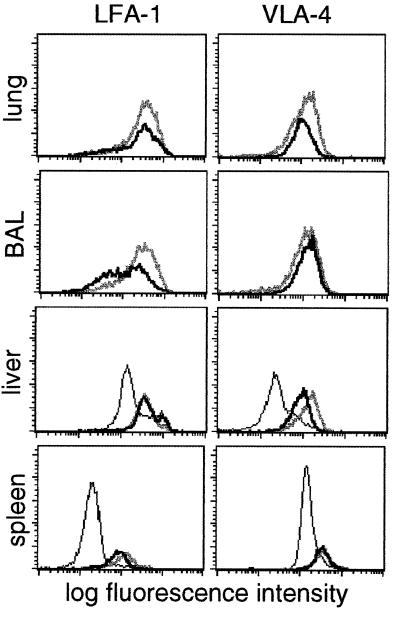
Figure 3.
Expression of LFA-1 and VLA-4 on CD8+ DbNP366+ T cells during secondary influenza infection. Flow-cytometric analysis of cells within a CD8+ lymphocyte gate isolated from lung, BAL, liver, and spleen is shown for total CD8+ T cells on day 0 (thin line), and for the CD8+ DbNP366+ set on day 8 (thick gray line) and day 13 (thick black line) after i.n. challenge of PR8-immune mice with the HKx31 virus.

