
Fig. 4.
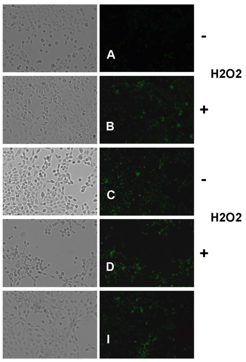

Anti-Apoptotic Effects of ECG and EGCG on H2O2-treated UROtsa Human Bladder Cells. Cells were treated with 40 μg/mL catechin agent or 1 mM Tiron in the presence or absence of 1 mM H2O2 and analyzed for apoptosis 24 h later using the Annexin V detection kit as described in Materials and methods. Annexin V-labeled apoptotic cells (green fluorescence) within the same microscopic field were viewed and imaged using bright field and fluorescence settings (FITC filter). For quantification, fluorescence was expressed as relative fluorescence units (RFU) per cell. (A, B) Negative control; (C, D) Tiron treatment; (E, F) ECG treatment; (G, H) EGCG treatment; (I) Positive (EtOH) control. Representative images are from three independent experiments.