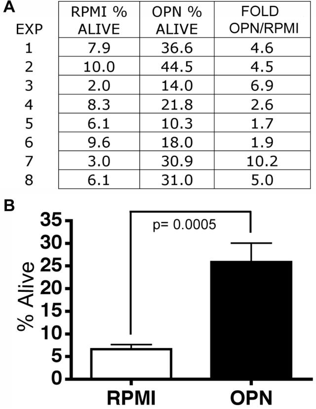
Figure 4. Osteopontin protects monocytes from apoptosis.

(A) In eight separate experiments (EXP 1-8), human monocytes were grown in non-adherent culture conditions for 24 hours. Shown is the percent of living cells (Annexin V and PI neg. cell population) in RPMI or cultured with 750 ng/ml of osteopontin (OPN). The fold is the increase in OPN compared to RPMI only. (B) The averages and standard error of the means are shown. OPN significantly increased the percentage of viable monocytes (4.7-fold, p= 0.0005).
