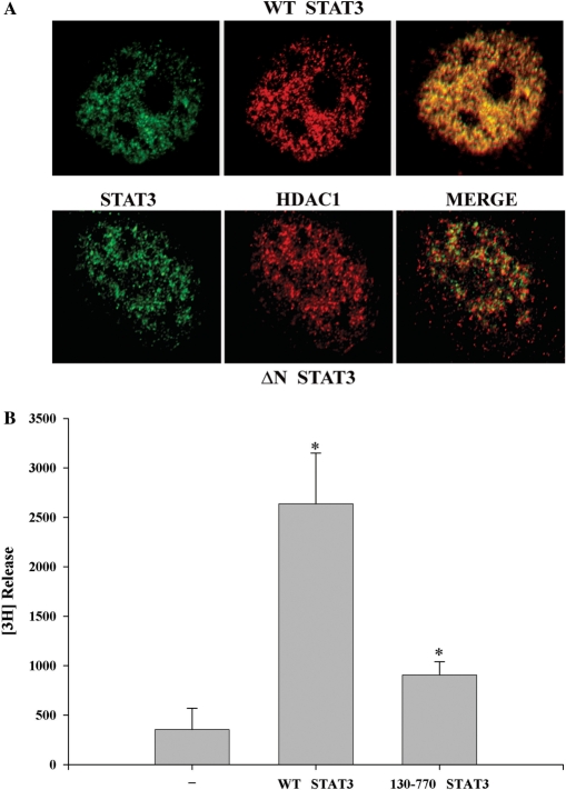
Figure 3.
(A) Localization of STAT3 with HDAC1. HepG2 cells were transfected with either V5-tagged WT STAT3 (1–770) (upper panel) or NH2-terminal deleted STAT3 (130–770) (lower panel) and stimulated with IL-6 for 30 min. Cells were fixed and incubated with anti-V5 Ab and antibody to HDAC1. Binding of primary antibody was detected by FITC- or Texas red-labeled secondary antibody. Confocal microscopy was performed on a Zeiss LSM510 META System using the 488 nm and 543 nm excitation for FITC and Texas red, respectively. Images were captured at a magnification of ×60. Co-localization was visualized (in the right panel, merge) by superimposition of green and red images using MetaMorph software. (B) Histone deacetylase activity is associated with STAT3 immune complex. HepG2 cells were transfected with either control empty vector, or full-length V5-tagged STAT3 or STAT3 NH2-terminal deleted mutant (aa 130–770) that weakly binds HDAC1. IL-6 stimulated WCEs were immunoprecipitated with anti-V5 Ab and these immune complexes were used for in vitro deacetylation assay to decaetylate [3H]-labeled histone H4 peptide (substrate), as described in the deacetylation assay kit protocol (UPSTATE Biochemicals). *P < 0.05 relative to empty vector control extract, t-test.

