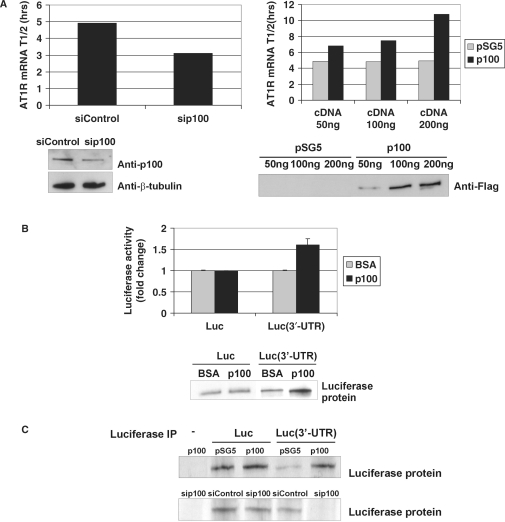
Figure 8.
p100 regulates positively AT1R mRNA stability and translation. (A) p100 silencing and overexpression. COS-1 cells were transfected with AT1R 3′-UTR construct with 30 nM of siControl or sip100 (left column) or with increasing amounts of pSG5 vector or p100-Flag (right column). The half-life of the AT1R mRNA in each p100 expression level condition was first plotted as linear fit and the resulting half-lives are shown in columns. Below are the control western blots for p100 silencing, β-tubulin expression and p100-Flag overexpression. (B) p100 enhances translation. In vitro translation level of luciferase coding region only (Luc) or of Luc(3′-UTR) transcript were compared in the presence or absence of affinity-purified p100. BSA was used as a control protein. Luciferase activity was measured from the in vitro translation reaction mixture. The results represent the means and SD of an average of three independent experiments. The mean luciferase value of the Luc transcript incubated with BSA was set to one. Biotin-labelled translation products were also detected by blotting with streptavidin-HRP antibodies (lower panel). (C) 35S incorporation. COS-1 cells were transfected with Luc or Luc(3′-UTR) constructs (except sample in the first lane) and co-transfected with pSG5 vector, p100-Flag expression plasmid, siControl or sip100, as indicated. After incubation with 35S-methionine, cells were lysed and subjected to IP with anti-luciferase antibody. The immunoprecipitated proteins were detected by autoradiography. Results are shown both after p100 overexpression (upper panel) and p100 silencing (lower panel).

