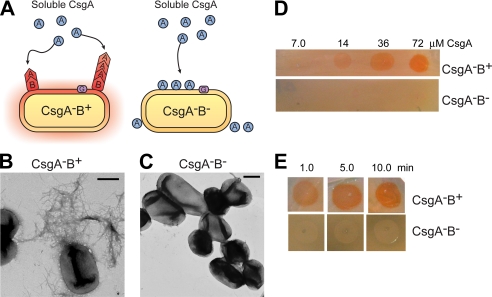
FIGURE 4.
Purified CsgA is efficiently nucleated when overlaid on CsgB-expressing cells. A, a schematic presentation of the overlay assay in which freshly purified and soluble CsgA is dripped onto cells expressing the nucleator protein, CsgB. In the presence of CsgB, CsgA (shown as a circle) undergoes a conformational change and polymerizes into a fiber (shown as a chevron). In the absence of CsgB, CsgA remains soluble and does not assemble into an amyloid fiber. B and C, negative-stain EM micrographs of CsgA-B+ (B) and CsgA-B- (C) cells grown on YESCA plates for 48 h at 26 °C that were overlaid with freshly purified 40 μm CsgA. Scale bars are equal to 500 nm. D, Congo red staining of CsgA-B+ and CsgA-B- cells after being overlaid with different concentrations of soluble CsgA. E, 72 μm CsgA was overlaid on CsgA-B+ and CsgA-B- cells and incubated for the indicated time intervals before staining for 5 min with Congo red solution.

