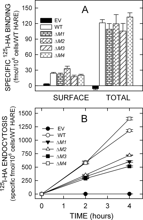
FIGURE 3.
125I-HA binding and endocytosis by cells expressing single motif 190-HARE CD mutants. Cells expressing EV, WT, or mutant 190-HARE were incubated in serum-free medium and washed, and specific 125I-HA binding at 4 °C or endocytosis at 37 °C was quantified as described under “Experimental Procedures.” A, cell surface and total cellular 125I-HA binding are indicated for cells expressing 190-HARE: WT (white bars), ΔM1 (horizontal rectangles), ΔM2 (diagonal rectangles), ΔM3 (diagonal lines), or ΔM4 (arrowheads) 190-HARE or EV (black). Values from 2 to 3 independent experiments are the means ± S.E. (n = 6–9) specific 125I-HA binding normalized for HARE expression relative to WT 190-HARE. B, specific 125I-HA internalization was determined at 37 °C for 2 and 4 h as described under “Experimental Procedures.” Values from 2 to 3 independent experiments are the mean ± S.E. (n = 6–9) specific 125I-HA endocytosis, normalized for HARE expression relative to WT. The plots show cells expressing WT (○), ΔM1 (▾), ΔM2 (Δ), ΔM3 (▪), or ΔM4 (□) 190-HARE, or EV (•).

