FIGURE 1.
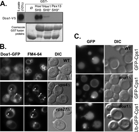
Association of Doa1 with the MVB sorting machinery. A, lysate from yeast expressing V5 epitope-tagged Doa1 was passed over beads bound with GST only (ø) or GST fused to the SH3 domain of Hse1 (Hse1 SH3), a mutant form of the SH3 domain (Hse1 SH3*), or the SH3 domain from Pex13 (Pex13 SH3). Also shown are the GST fusion proteins used in this analysis. B, Doa1-GFP (expressed from pPL3327) was localized in wild type cells (WT), vps4Δ cells, and vps27Δ cells. Cells were counterlabeled with the endocytic tracer dye FM4-64. C, GFP-Cps1 was correctly localized to the vacuole lumen in wild type cells but not doa1Δ mutant cells, the latter of which showed accumulation of GFP-Cps1 at the limiting membrane of the vacuole. The lower panel shows sorting of Ub-GFP-Cps1 to the vacuole lumen in doa1Δ cells. Also shown are corresponding DIC images.

