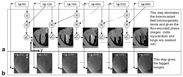
Figure 1.
Diagram representing the postprocessing steps used to reconstruct the tagged images and the chamber blood flow images from the reconstructed complex SPAMM n' EGGS images I(p,t). A: A sliding-window phase-sensitive reconstruction (un-encoded images used as phase reference) used to isolate the blood flow velocity terms. B: The tagged images obtained from magnitude reconstruction.

