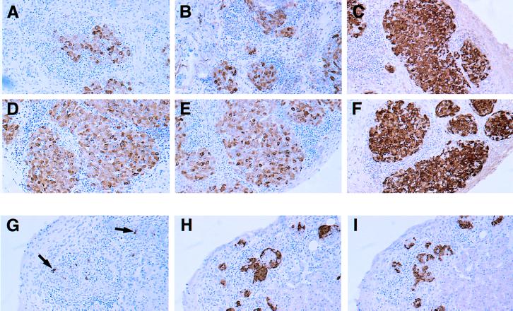
Figure 2.

Histopathology of NOD or NODlpr islet grafts in diabetic NOD mice. Two examples of NOD (A and B) and NODlpr (D and E) islet grafts are shown at 21 or 15 days after transfer of diabetogenic spleen cells. The grafts from NOD mice that received nondiabetic spleen cells also are shown: NOD (C) and NODlpr (F) 61 days after transfer. (G–I) Appearance of NODlpr fetal pancreas islet grafts 30 days after being grafted under the kidney capsule of spontaneously diabetic female recipients. Sections were analyzed by immunohistochemistry (brown color) for insulin (A–G), somatostatin (H), and glucagon (I) and were counter-stained with haematoxylin. Arrows in G indicate occasional remaining β cells. (×100.)
