
Figure 3.1.
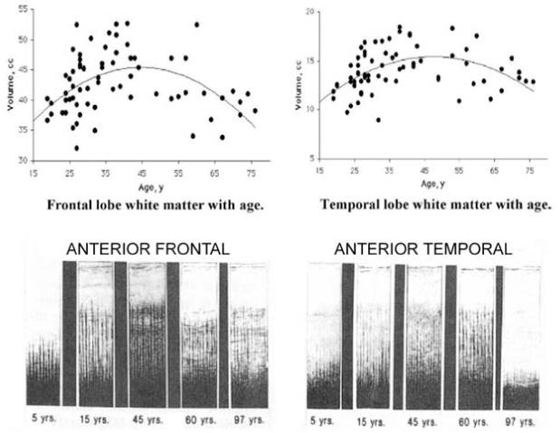
Quadratic trajectories of myelination of human brain over the life span. Myelination (Y axis) versus age (X axis) in frontal (left) and temporal (right) lobes of normal individuals. Top figures are in vivo data from Bartzokis et al. (2001). Lower figures show postmortem intracortical myelin stain data from Kaes (1907) adapted and reproduced in Kemper (1994). Used with permission.
The data were acquired 100 years apart, yet the two samples of normal individuals show remarkably similar myelination trajectories in the two regions. Note that different brain regions have significantly different myelination trajectories even when the regions are similar, as is the case with these two association regions. Peak myelination is reached in the frontal lobe at age 45 and even later in the temporal lobe.