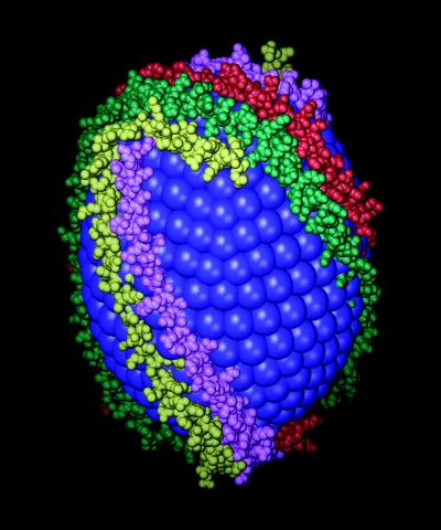
Figure 5.
Hypothetical model of apo A-I bound to spherical HDL. Two dimers of apo Δ(1–43)A-I are shown as CPK models, colored as in Fig. 4. The lipid head groups are represented by blue balls. At the top of the model are helices A10, D5, C5, and B10 (left to right). Separation of helices A10 and B10 of the A/B dimer allows the C/D dimer to pass through the gap, over the top of the sphere.

