
Figure 2a. BAFF expression on CD14+ monocytes of CVID subjects and controls.
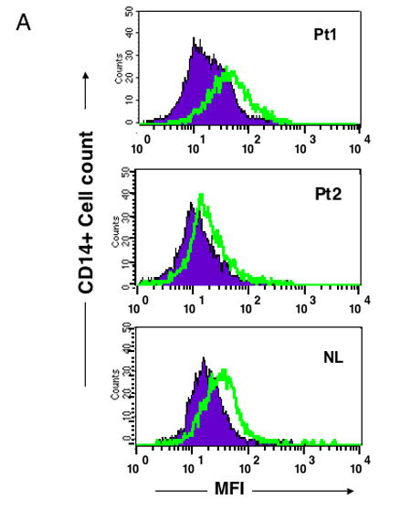
BAFF expression was demonstrated by mean fluorescence intensity (MFI) as detected by FACScan gating on CD14+ peripheral blood monocytes from freshly isolated PBMC from two representative CVID subjects shown here (of 10 tested) in comparison to a normal control (of 10) done at the same time. CD14+ cells were identified by FITC labeled anti-CD14 antibody; BAFF was detected by a rat monoclonal anti-BAFF followed by a goat anti rat CY5 conjugate, using appropriate rat IgG2a isotype controls. The closed areas indicate isotype controls and open areas, BAFF staining.