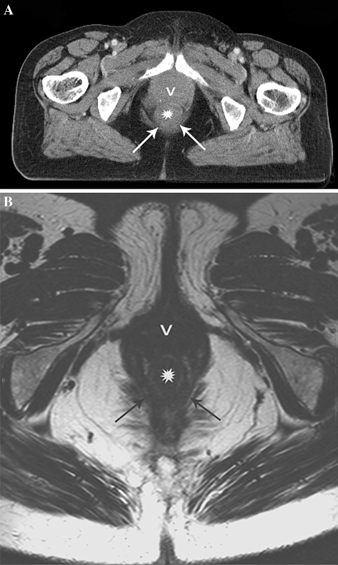
Fig. 4.
Normal rectal wall staged as tumor invasion of the MRF on CT due to insufficient anatomical detail. A Axial MS-CT image suggest a thickened rectal wall interpreted as distal tumor (asterisk) contacting the pelvic floor (arrows) and vagina (v). B Axial T2-weighted MR image at the same level clearly depicts a normal rectal wall (asterisk) as well as surrounding anatomy (arrows = pelvic floor; v = vagina).

