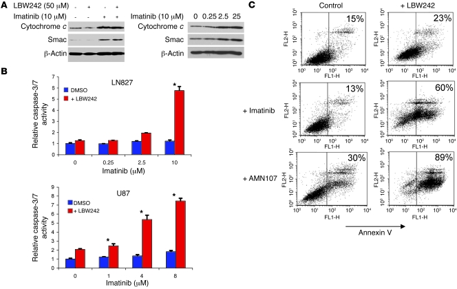
Figure 3. PDGFR and IAP inhibition combine to enhance caspase activity and activate apoptosis in glioma cells.
(A) LN827 cells were treated with drugs as indicated for 48 hours, following which the cellular cytoplasm was separated from mitochondria. The cytosol was collected and subjected to immunoblotting for cytochrome c and Smac/DIABLO. (B) LN827 and U87 cells were treated with imatinib at the dosages indicated for 48 hours in combination with LBW242 (50 μM) or DMSO control. Caspase-3/7 activity is expressed relative to controls as mean ± SEM of triplicates. *P < 0.01, 2-tailed Student’s t test. (C) Apoptosis was measured as the proportion of cells staining positive for annexin V after 72 hours of incubation with LBW242 (50 μM) in combination with or without imatinib (10 μM) or AMN107 (5 μM). Cells to the left of the divider in each panel are negative for annexin V, and positive cells are to the right. The number in the upper right corners indicates the percentage of annexin V–positive cells in each treatment group. Similar results were obtained in 3 independent experiments.

