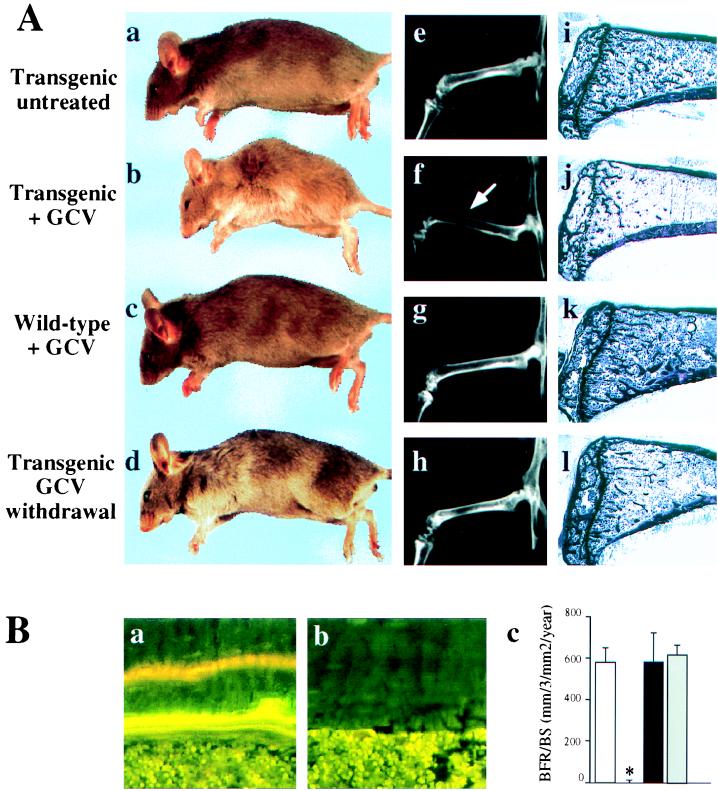
Figure 2.

Bone-formation parameters in GCV-treated animals. (A) Morphological, radiological, and histological comparison of 10-week-old tg mice untreated (a, e, and i) or treated daily for 4 weeks with GCV (b, f, and j), 10 week-old wt mice treated daily for 4 weeks with GCV (c, g, and k), or 14 week-old treated tg mice 4 weeks after GCV withdrawal (d, h, and l). Note the thinning of the cortices (arrow) and the lucent aspect of the bones on the x-rays (f) as well as the loss of the trabecular bone (j) in GCV-treated tg mice. These abnormalities were reversible after GCV withdrawal (compare f with h and j with l). (B) Dynamic histomorphometric analysis after tetracycline/calcein double labeling. Fluorescent micrographs of the two labeled mineralization fronts in representative section at the mid diaphysis of the tibia of wt (a) or tg mice (b) at the end of a 4-week GCV treatment period. (c) Measurement of the bone formation rate. Open bar, 10-week-old untreated tg mice; hatched bar, 10-week-old tg mice GCV-treated for 4 weeks; closed bar, 10-week-old wt mice GCV-treated for 4 weeks; gray bar, 14-week-old tg mice 4 weeks after GCV withdrawal. The asterisk indicates a statistically significant difference between wt and tg mice (P < 0.001; n = 4).
