Figure 1.

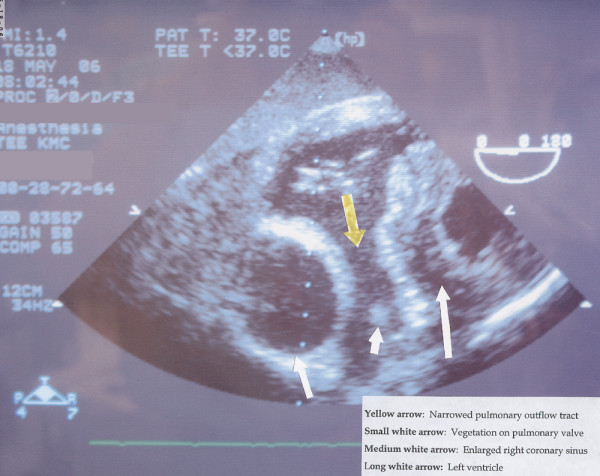
A transesophageal echocardiogram depicting an enlarged right coronary sinus (medium white arrow) and identification of the vegetation on the pulmonary valve (small white arrow).

A transesophageal echocardiogram depicting an enlarged right coronary sinus (medium white arrow) and identification of the vegetation on the pulmonary valve (small white arrow).