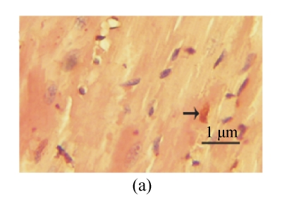
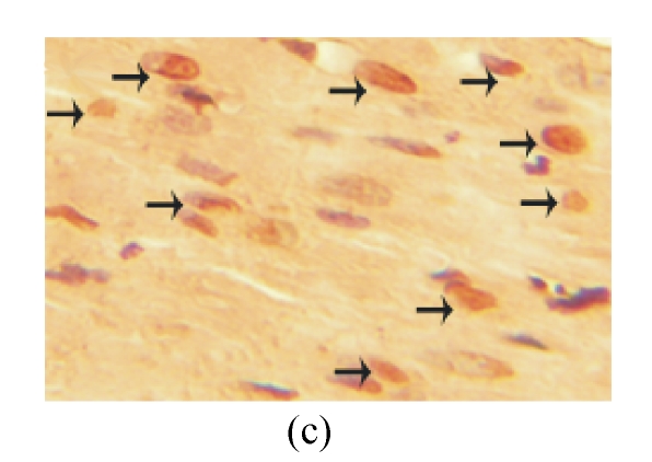
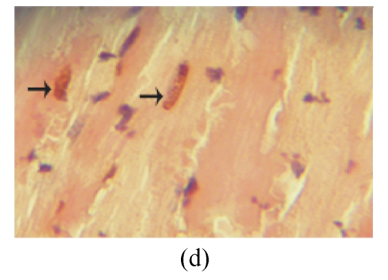
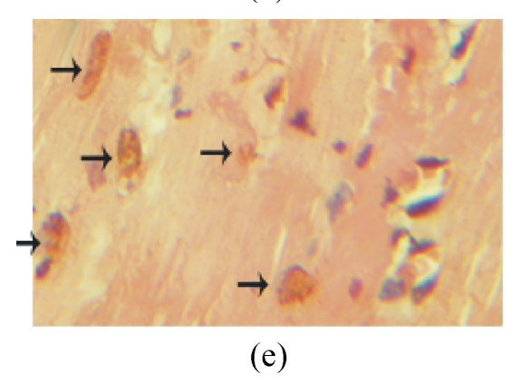
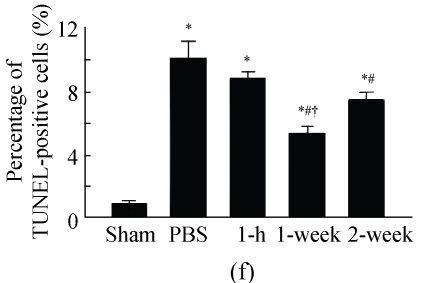
Fig. 3.
The apoptosis of cardiomyocytes was tested by TUNEL assay. (a) Sham-operated group; (b) PBS group; (c) 1-h group; (d) 1-week group; (e) 2-week group; (f) Percentage of TUNEL-positive cells. The percentage of apoptotic cells was determined in 5 random microscopic fields totally at least 1000 cells/section. A total of 20 sections were analyzed for each rat from the 5 groups. * P<0.01 vs Sham group; # P<0.01 vs PBS group; † P<0.05 vs 1-h and 2-week groups. The black arrows indicate the apoptotic cells (brown nucleus staining)

