Fig. 7.
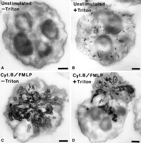
Electron micrographs of thick sections (0.35 μm thickness) of human neutrophils incubated for the cytochemical detection of alkaline phosphatase. a An unstimulated cell that was incubated for the detection of alkaline phosphatase activity in the absence of cell permeabilization with Triton X-100. Note that there is no reaction in the absence of permeabilization. b A cell that was incubated for the detection of alkaline phosphatase activity following cell permeabilization. In this case, numerous small rod-shaped organelles containing reaction product are evident. c A cell stimulated with f-Met-Leu-Phe (FMLP) and cytochalasin B (Cyt.B) that was incubated for the detection of alkaline phosphatase activity in the absence of cell-permeabilization. Note the dramatic rearrangement of the alkaline phosphatase compartments into elaborate tubular structures. These tubular structures appear to be in continuity with the extracellular space since permeabilization was not required to detect the enzyme activity. d A cell stimulated with f-Met-Leu-Phe (FMLP) and cytochalasin B (Cyt.B) that was incubated for the detection of alkaline phosphatase activity in the presence of cell permeabilization. This cell has a appearance similar to the cell treated in the same manner but without permeabilization. The cytochemical reaction was carried out as we have described previously (Kobayashi and Robinson 1991). Bars: 1 μm

