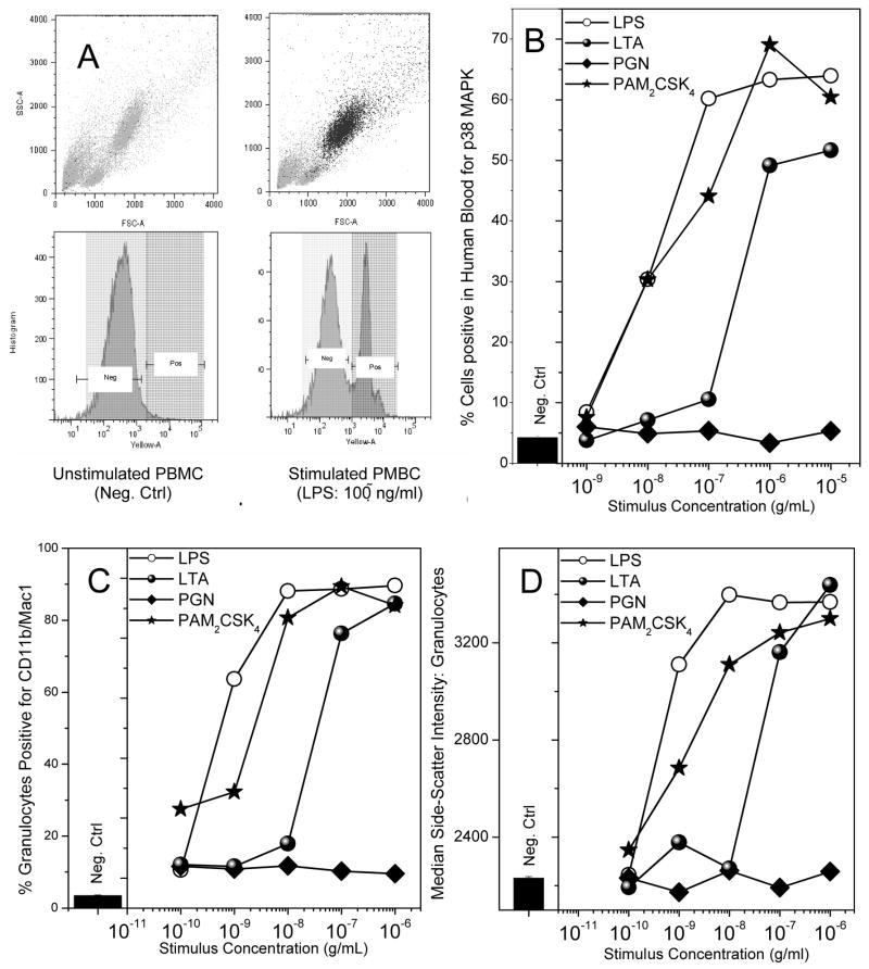
Fig. 4.
(A) Depiction of negative control (left; medium alone) and positive control (right; LPS-treated) flow cytometric forward/side scatter and p38 MAPK abundance histogram plots of human blood samples. A marked increase in p38 MAPK signal was observed in the granulocyte population. (B) p38 MAPK phosphorylation in whole human blood treated with 1:10 serial dilutions of the ligands. Blood samples were exposed to stimuli for 15 minutes, and cells were fixed, permeabilized, stained with anti-p38 MAPK antibody-PE conjugate, and analyzed by flow cytometry. (C) Up-regulation of the surface marker CD11b in granulocytes upon treatment with the various ligands of interest. Whole human blood was treated with the ligands under investigation, serially diluted 1:10, for 1 hour. Cells were then stained with anti-CD11b/Mac-1 antibody for 30 minutes on ice, fixed, washed, and analyzed via flow cytometry. (D) Dose-dependent increase in median side-scatter intensity of the granulocyte population in human blood treated with the ligands of interest was carried out on the samples from (C). A robust increase in the median-side scatter intensity, corresponding to increase in cellular granularity, in those samples which were stimulated with LPS and Pam2CSK4 was observed. Representative results from three independent experiments performed on blood samples from different donors are shown in B, C and D.

