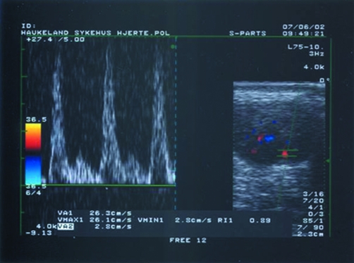
Figure 3 Colour Doppler image of a transverse section of Achilles tendon showing intratendinous blood vessels (right hand frame) and power Doppler (left hand frame) assessment of intratendinous blood flow velocity in systolic peak (26.1 cm/s) and diastolic end (2.8 cm/s), giving a resistive index of 0.89, indicating minor inflammation.

An official website of the United States government
Here's how you know
Official websites use .gov
A
.gov website belongs to an official
government organization in the United States.
Secure .gov websites use HTTPS
A lock (
) or https:// means you've safely
connected to the .gov website. Share sensitive
information only on official, secure websites.
