Figure 1.
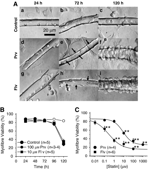
Pravastatin and fluvastatin induced vacuolation and cell death in FDB fibres. (A) Phase contrast micrographs. (a–c) A control myofibre after 24 (a), 72 (b) and 120 h (c) in culture with fibroblasts in the background. (d–f) A myofibre cultured with 100 μM pravastatin (Prv) for 24 (d), 72 (e) and 120 h (f). Note that the background cells remained intact whereas damage was prominent in the skeletal myofibre. (g–i) A myofibre cultured with 10 μM fluvastatin (Flv) for 24 (g), 72 (h) and 120 h (i). Note that the background cells remained intact; whereas there was prominent damage in the skeletal myofibre. Unlike pravastatin treatment, the background cells disappeared after Flv treatment. Arrows indicate vacuoles induced by the statin treatment (e, h). (B) Time-dependent changes in the percentage of Trypan blue positive myofibres among control, pravastatin- and fluvastatin-treated cultures. (C) Comparison of cell death induced by Prv and Flv. Myofibres were incubated with various concentrations of Prv (closed circles) or Flv (closed squares) for 120 h. LC50 values for Prv and Flv are 8.6 and 0.3 μM, respectively (**P<0.01, compared with the control).

