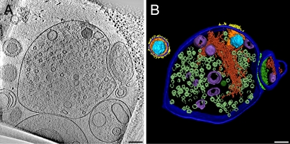
Fig. 2.
HSV-1 enters the presynaptic part of synaptosomes. (A) A 16-nm-thick slice of a tomogram from a synaptosome inoculated with HSV-1 for 60 min at 25°C. A capsid is localized inside the presynaptic element on the left characterized by abundant synaptic vesicles. A smaller postsynaptic fraction (right) is still attached via adhesion molecules to the synaptic cleft. Glycoprotein spikes are visible on the outer phase; tegument proteins correspond to the local densities near the cytoplasmic phase of the plasma membrane. Note structural changes between the entire virion (upper left corner) and the one that had entered the synaptosome. (B) Surface rendering of one virion and the synaptosome from the tomogram in A: capsid (light blue), tegument (orange), glycoproteins (yellow), cell membrane/viral membrane (dark blue), actin (dark red), vesicles (purple), synaptic vesicles (only partially segmented—metallic green), synaptic cleft (light green), and postsynaptic density (green). (Scale bars, 100 nm.)

