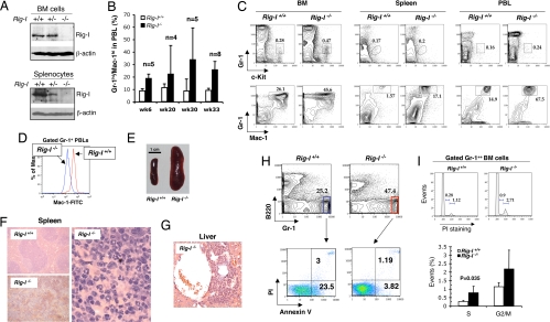
Fig. 2.
Rig-I deficiency leads to a progressive granulocytosis. (A) Rig-I expression levels within whole BM cell and spleen cell lysates from Rig-I+/+, Rig-I+/−, or Rig-I−/− mice were measured by Western blotting assay. (B) Onset and progress of the abnormal Gr-1hi/Mac-1hi granulocytic production in PBLs (mean ± SD). (C) Representative results of analyzing the percentages of c-Kit+Gr-1lo granulocytic progenitors (GPs) and Gr-1++Mac-1++ granulocytes in BM, spleen, and PBLs of Rig-I+/+ or Rig-I−/− mice at the age of 6 months. Notably, Mac-1 signal strength of Rig-I−/− myeloid cells is lower than that in wild-type counterparts. (D) Comparison of Mac-1 expression strengths in Rig-I+/+ and Rig-I−/− peripheral blood granulocytes. (E) Rig-I deficiency resulted in severe splenomegaly in aged mice. A representative enlarged spleen frequently found in old Rig-I−/− mice (≥9 months) is shown. (F) Hematoxylin-eosin staining of splenic sections shows the disrupted splenic nodules by the infiltrating granulocytic cells (Right and Lower Left) in Rig-I−/− mice in comparison with the normal splenic nodular structure. (G) Rig-I−/− granulocytes invaded hepatic tissues. An infiltration focus of Rig-I−/− granulocytes into the perivascular compartment of liver is shown. (H) Rig-I−/− granulocytes survived better than their normal counterparts. The freshly isolated primary BM cells were simultaneously stained with Gr-1, B220, Annexin V, and propidium iodide (PI). One set of representative flow cytometric data of gated B220− Gr-1++ myeloid cells from Rig-I−/− or Rig-I+/+ mice is shown. (I) Cell cycle statuses of Gr-1++ BM myeloid cells were measured by PI staining after permeabilization. The average lengths for indicated cell cycling phases are expressed as mean ± SD (n = 5, S phase P < 0.05).

