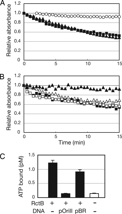
Fig. 3.
Hydrolysis of ATP by RctB and DnaA. (A) Spectrophotometric assay of generation of ADP after incubation of 10 μM DnaA (■), 10 μM RctB (▲), or no protein (○) with ATP. (B) Influence of pOriCII DNA (triangles) or pOriCI DNA (squares) on ATP hydrolysis by DnaA (open symbols) or RctB (closed symbols). (C) Inhibition of RctB binding of ATP by pOriCII DNA. A total of 80 nM of RctB was incubated with 1 nM of P32-αATP in the presence or absence of 180 ng of pBR or pOriCII DNA. A control experiment without RctB and DNA shows the background level of P32-αATP binding to the filter. The results shown in A and B are representative of at least three experiments, and the data in C represent the average and standard deviation derived from three experiments.

