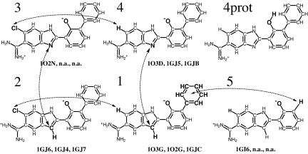
Fig. 1.
Inhibitors of serine proteases from ref. 37 and 38 used as a test system. Arrows show simulated mutations. Note that four of the mutations form a closed cycle, 1 → 2 → 3 → 4 → 1. The PDB codes (40) of the corresponding complexes with trypsin, thrombin, and uPA are also shown if they exist. According to ref. 37, the 1GJC complex contains ligand 1, but the 1GJC files from the PDB contain ligand 4. Thus, the complex of uPA with ligand 1 was obtained by substituting the corresponding nitrogen of ligand 4 by a CH group without change of coordinates of any other atoms. Ligands are shown as they are in complexes: with deprotonated phenyl oxygen (38). In solution the ligands will be largely protonated at this site as shown for ligand 4 in the upper right corner.

