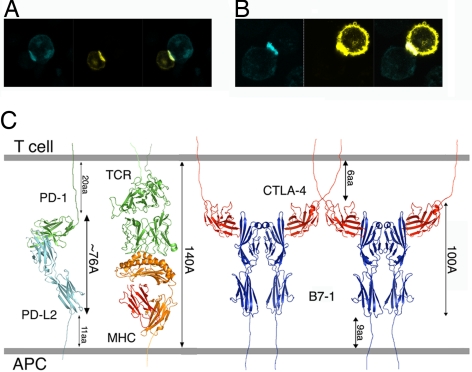
Fig. 6.
PD-1/PD-L interaction at the cell–cell interface. Noncovalent interactions between PD-1 and PD-Ls are sufficient to drive their enrichment at a pseudosynapse. (A and B) PD-1 and PD-L1 (A) or PD-L2 (B) expressed in CHO cells are recruited to the cell–cell contact area and form conjugates that are analogous to the immunological synapse. (Left) PD-1-CFP-expressing cells in blue. (Center) PD-L1-YFP or PD-L2-YFP-expressing cells in yellow. (Right) Overlay of the CFP and YFP images. (C) Model of the PD-1/PD-L2 complex in the immunological synapse. A number of receptor–ligand assemblies have dimensions that are compatible with colocalization to the central zone of the immunological synapse.

