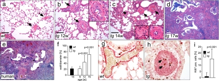
Fig. 2.
Obliteration of pulmonary arteries precedes pulmonary fibrosis in fra-2tg mice. (a–d) Time course analysis of pulmonary disease in fra-2tg mice between weeks 12 and 17 identifying obliteration of pulmonary arteries as early event of pulmonary disease in transgenic mice (arrows in b and c; magnification in b; right magnification in c; n > 5 per time point). In contrast, pulmonary arteries in wild-type controls appeared normal (arrows in a). Arteriostenosis was followed by inflammation and the appearance of fibroblastic proliferations (arrowhead and left magnification in c). (d and e) The histological alterations at later stages resembled human NSIP- and IPF-related usual interstitial pneumonia. (Scale bars: 200 μm.) (e) A sample from a patient with IPF. The short arrow indicates an occluded vessel. (f) Quantification of arteriostenosis. Thickness of the vessel walls is given as a percentage of the vessel diameter (n ≥ 15 vessels from at least three mice per time point). (g and h) Dual immunohistochemistry for α smooth muscle actin (αSMA) (red) and Ki67 (brown) at 14 weeks of age for wild-type (g) and fra-2tg (h) mice. Arteriostenosis was caused by neointima formation due to proliferation of αSMA-expressing VSMCs (h, arrows). (i) Proliferation in the vascular wall was quantified by immunohistochemistry for Ki67. The amount of Ki67-positive nuclei is given in percentage (n = 20 vessels from two mice per genotype at 14 weeks of age).

