Fig. 5.
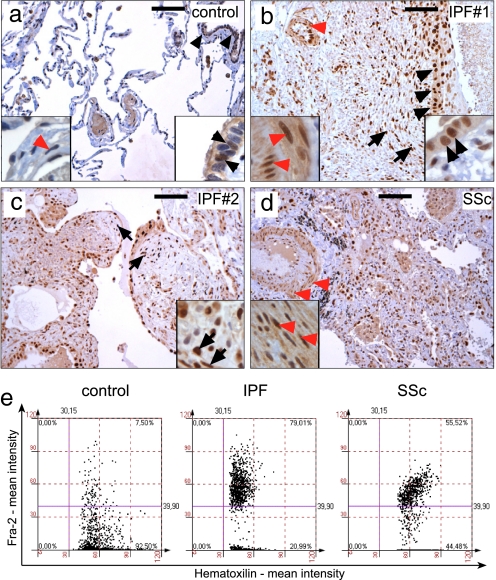
Fra-2 is strongly expressed in human pulmonary fibrosis as determined by immunohistochemistry. (a) In control samples, most nuclei were negative for Fra-2 (blue-stained nuclei). Nuclear localization of Fra-2 (brown-stained nuclei) was detected in some bronchial epithelial cells (black arrowheads, right magnification), whereas almost no expression was observed in VSMCs (red arrowheads, left magnification). (b and c) Strong nuclear localization of Fra-2 in two representative samples from patients diagnosed as IPF (a comparable signal for Fra-2 was observed in all IPF samples). Expression of Fra-2 was strongest in bronchial and pneumocytic epithelia (black arrowheads, right magnification in b), fibroblastic foci (short arrows in b and c, magnification in c), endothelial cells and VSMCs of pulmonary arteries (red arrowheads and left magnification in b). (d) Fra-2 expression in lung samples of immunologically determined systemic sclerosis (SSc). Note the vascular remodeling of pulmonary arteries with prominent Fra-2 staining in VSMCs (red arrowheads, magnification). (Scale bars: 100 μm.) (e) Tissue cytometric analysis in control, IPF, and systemic sclerosis samples confirmed up-regulation of Fra-2 expression. Representative examples are shown. Fra-2-negative nuclei and Fra-2-positive nuclei were counted, and the signal intensity was quantified by using HistoQuest software (TissueGnostics). Four randomly chosen fields on control, IPF, and systemic sclerosis sections were quantified and are shown as merged images.

