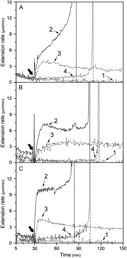
Figure 5.
Enhancement of reconstituted expansin-induced extension of growing cell wall by recombinant Pel1, PelA, and PG. A, Enhancement of Pel1. Reconstituted expansin-induced extension of apical segments of cucumber hypocotyls following replacement of bathing buffer alone (line 1), or containing 1 mg mL−1 of expansin plus 0.44 units mL−1 of Pel1 (line 2, representative), or containing 1 mg mL−1 of expansin (line 3, mean), or containing 0.44 units mL−1 of Pel1 (line 4, representative). B, Enhancement of PelA. Reconstituted expansin-induced extension of apical segments of cucumber hypocotyls following replacement of bathing buffer alone (line 1, mean), or containing 1 mg mL−1 of expansin plus 6.10 units mL−1 of PelA (line 2, representative), or containing 1 mg mL−1 of expansin (line 3, mean), or containing 6.10 units mL−1 of PelA (line 4, representative). C, Enhancement of PG. The reconstituted expansin-induced extension of apical segments of cucumber hypocotyls following replacement of bathing buffer alone (line 1, mean) or containing 1 mg mL−1 of expansin plus 1.0 units mL−1 of PG (line 2, representative), or containing 1 mg mL−1 of expansin (line 3, mean), or containing 1.0 units mL−1 of PG (line 4, representative). Details are described in Figure 1A. Curves are the mean (mean) or representative (representative, based on its breakage) of three independent experiments. Thick arrows indicate when bathing buffer was replaced.

