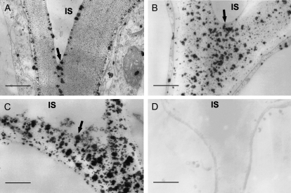
Figure 8.
Calcium localization in cell walls along the growth gradient of cucumber hypocotyls. A to C, Transverse sections were made in the region of 0.5 cm (A), 4 cm (B), and 8 cm (C) below the hook and assayed with the potassium pyroantimonate method for calcium localization. The particles of calcium precipitation in cell walls of the cell junctions between epidermis and subepidermis were displayed. Particles of calcium precipitation disappeared in the walls of basal segments that had been pretreated with 100 mm EGTA. D, Details are described in Fig. 6. The experiment was repeated three times with the same results. Three cucumber hypocotyls and nine transverse sections from each region of each hypocotyl were examined in each experiment. IS, intercellular space. Bars = 0.5 μm.

