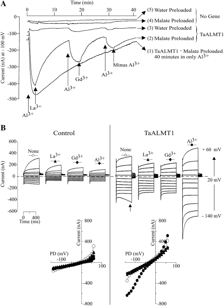
Figure 2.
Cation specificity of TaALMT1 activation by extracellular Al3+. A, Extracellular Al3+ is required to maintain TaALMT1 activation. Trace 1 shows the TaALMT1-mediated inward current activity over a 40-min period following activation by Al3+ (100 μm AlCl3 concentration at the time indicated by the arrow) in a malate-preloaded cell. Traces 2 and 3 show the activity of the TaALMT1 inward current in malate-preloaded (trace 2) or water-preloaded (trace 3) cells following activation by Al3+ and subsequent exchange of solutions containing 100 μm of the trivalent cations indicated at the bottom of the traces. Recordings from control cells preloaded with malate or water and subjected to identical treatments as those shown for traces 2 and 3 are shown for reference (traces 4 and 5, respectively). All traces were taken from different cells bathed in ND96 solution. Similar responses were observed for at least three other cells for each case and ionic condition. B, Examples of families of currents recorded from control (left) and TaALMT1-expressing (right) cells preloaded with malate and bathed in ND96 solution containing 100 μm lanthanide (described at the top of each set of traces). Currents shown are in response to voltage pulses ranging from −140 to +60 mV. The arrow in the right panel indicates the endogenous time-dependent inward currents recorded at large negative potentials occasionally detected in some cell batches. Note that the presence of this endogenous current was only evident when Al3+ or analog lanthanides were not present in the bath solution. Similar recordings were observed in a total of six control and six TaALMT1-expressing cells tested. The bottom panel shows the I/V relationships obtained from the above recordings, with the symbols corresponding to those shown at the top of each set of traces.

