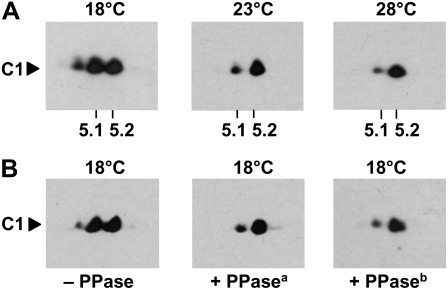
Figure 2.
C1 is hyperphosphorylated at the low temperature. A, C1 immunodetection from proteins of crude extracts separated by 2-DE. Cells were grown at 18°C, 23°C, and 28°C and harvested during early day (LD2). Crude extracts were prepared, and proteins (300 μg per assay) were separated by standardized 2-DE (see “Materials and Methods”). For the first dimension, an immobilized pH gradient strip of pH 3 to 10 was taken, and in the second dimension, 10% SDS-PAGE was used along with a molecular mass standard. The proteins were then immunoblotted with anti-C1 antibodies. The positions of pH 5.1 and 5.2 that are close to the theoretical pI of C1 (5.17) are indicated. B, The same procedure described for A was undertaken with cells grown at 18°C (− PPase). In one case (+ PPasea), the extract was treated for 30 min with λ PPase (New England Biolabs) at 30°C according to the protocol of the supplier. In another case (+ PPaseb), the amount of PPase used was increased five times.

