Figure 3.
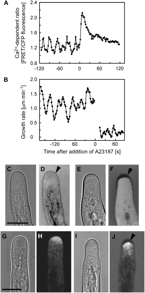
Effects of the Ca2+ ionophore A23187 on cytosolic Ca2+ and growth of Arabidopsis root hairs. A, Treatment with 10 μm A23187 triggers a rapid increase of cytosolic Ca2+ in a growing root hair. Ca2+ levels were measured in the approximately 30-μm2 ROI at the root hair apex indicated in Figure 1A. Representative results of 10 measurements are shown. B, Treatment with 10 μm A23187 arrests root hair tip growth. All detectable growth of the root hair had ceased by 14 s, when measurements resumed. The nongrowing root hair did not remain in the same plane as the root axis continued to shift after treatment, necessitating constant refocusing. At or below the limits of resolution for the tracker (0.2 μm min−1), this combination of changes in focal plane and root expansion appear as very slow growth for this example. Representative results of 10 measurements are shown. C to F, Bright-field and fluorescence images of root hairs loaded with fluorescein diacetate and immersed in medium containing fluorescein dextran. C and D, Untreated growing control root hairs. Cell wall (arrowhead) thickness is indicated by the exclusion of cytosolic and extracellular fluorescein fluorescence. E and F, A23187-induced Ca2+ increase and growth inhibition are accompanied by a thickening of the apical root hair cell wall (arrowhead). Images were acquired 1 h after the start of ionophore treatment. Representative results of 11 measurements are shown. G to J, Bright-field and fluorescence images of root hairs expressing YFP-RabA4b. G and H, Untreated growing control root hairs. Note that YFP-RabA4b accumulates at the apex of the growing root hair. I and J, A23187-induced apical cell wall thickening is accompanied by an accumulation of YFP-RabA4b at the cell apex. The root hair was immersed in medium containing fluorescein dextran to help visualize cell wall thickness (arrowhead). Representative results of 10 measurements are shown. Bars = 10 μm.

