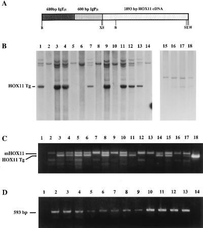
Figure 1.
Structure and detection of the IgHμ-HOX11 transgene. (A) Diagrammatic representation of the 3.18-kb HOX11 transgene consisting of the human HOX11 cDNA under the transcriptional control of the murine IgH promoter (IgPμ) and enhancer (IgEμ). Restriction sites: B, BglII; X, XbaI; S, SmaI; E, EcoRI; H, HindIII. (B) Southern blot showing mouse genomic DNA from nontransgenic and transgenic mice from HOX11 mouse lines C2 (lanes 1–14) and D11 (lanes 15–18) digested with BamHI or XhoI/NotI, respectively, and hybridized with either the HOX11 cDNA (lanes 1–14) or a 1.4-kb 5′ HOX11 genomic probe (lanes 15–18). The HOX11 transgene (HOX11 Tg) is indicated. (C) PCR analysis used to distinguish control and HOX11 mice from the C5 founder line. The primers amplify both endogenous murine (mHOX11) and a 499-bp HOX11 transgenic (HOX11 Tg) fragment. Lane 1 contained a negative control, and lanes 2–15 contain amplified DNA from transgenic and nontransgenic mice followed by CD-1 nontransgenic DNA, positive transgenic DNA, and a plasmid containing the entire HOX11 cDNA. (D) Reverse transcription–PCR analysis showing expression of the HOX11 transgene in hematopoietic tissues and tumors from C2 (lanes 9, 11, and 12), C5 (lanes 6 and 10), and D11 (lanes 3–5, 7, 8, and 13) HOX11 mice affected with lymphoma or myeloid hyperplasia. K3P (lane 2), used as a positive control, is a t(10;14) positive human T-ALL cell line (5). Lanes 3–13 contain the 593-bp fragment amplified from cDNA generated from RNA isolated from the indicated tissues: thymus (lanes 3, 7, and 8), kidney (lanes 4 and 6), marrow (lane 5), spleen (lanes 9 and 11), tumor (lanes 10, 12, and 13).

