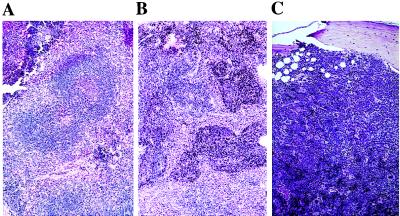
Figure 2.

Lymphoid and myeloid hyperplasia in young HOX11 transgenic mouse tissue sections stained with hematoxylin and eosin. (A) Section of a spleen from a 4-week-old mouse showing fusion of germinal centers. (B) Section of a spleen from a 6-month-old transgenic mouse showing lymphoid hyperplasia with characteristic invasion of red pulp areas. (C) Myeloid hyperplasia in the bone marrow of a 6-month-old HOX11 transgenic mouse. Note the increased cellularity and loss of fatty spaces, but all hematopoietic cell types are present. (Magnification: ×40.)
