
Figure 1.
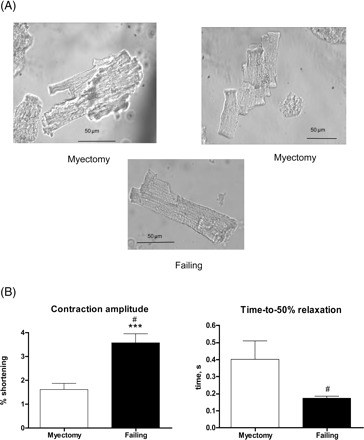
Contraction of isolated myocytes from myectomy muscle. (A) Two ventricular myocytes from myectomy samples, showing branched appearance, compared with the more usual rod-shaped appearance of a ventricular myocyte from failing heart. (B) Contraction amplitude (% shortening) and time-to-50% relaxation of ventricular myocytes from myectomy (21 myocytes/10 hearts) and failing (41 myocytes/16 hearts) samples. #P < 0.001, variances significantly different; ***P < 0.001, means significantly different.