Figure 4.
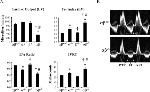
Evidence for cardiac failure in PGC-1αβ-deficient mice. To evaluate cardiac function noninvasively in all four genotypes, high-resolution echocardiography was performed within a few hours after birth. (A) Bar graphs show representative indices of systolic (cardiac output), diastolic (E/A ratio, IVRT), and combined (Tei index) left ventricular performance. (B) Representative images of the trans-mitral/left ventricular outflow tract (LVOT) Doppler spectra from wild-type (αβ+/+) and PGC-1αβ−/− (αβ−/−) mice demonstrate markedly altered cardiac time intervals and reduced LVOT velocities in the double null mice. (IVRT) Isovolumic relaxation time; (IVCT) isovolumic contraction time; (ET) ejection time.

