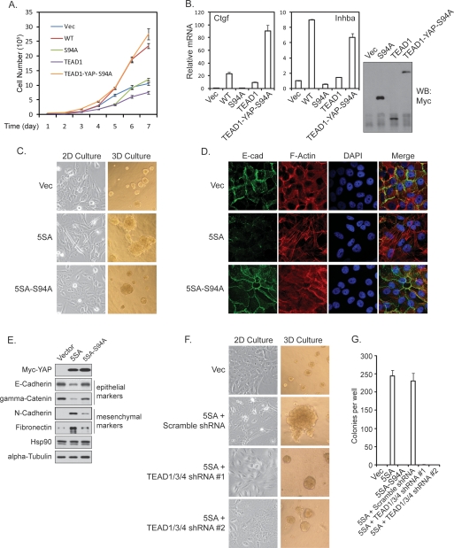
Figure 2.
TEAD is required for YAP activity in growth promotion and EMT. (A) YAP-S94A is defective in promoting cell growth. The growth curve of NIH-3T3 stable cells with expression of Vector, YAP, YAP-S94A, TEAD1, or TEAD1-YAP-S94A was determined. (B) Fusion of YAP-S94A with TEAD1 rescued YAP target gene expression. (Right panel) NIH-3T3 stable cells with expression of YAP-S94A, TEAD1, and TEAD1-YAP-S94A fusion protein were generated, and the expression of these proteins was shown by anti-Myc-tag Western blot. The expression of Ctgf and Inhba, two YAP target genes in NIH-3T3 cells, was measured by quantitative PCR. The induction of these two genes by YAP-WT was also shown for comparison. (C) YAP-5SA-S94A is compromised in eliciting EMT-like morphology. Indicated MCF10A stable cells were cultured in monolayer or in 3D on reconstituted basement membrane for 16 d before pictures were taken. (D) YAP-5SA-S94A is defective in reducing membrane E-cadherin and cortical actin. The indicated MCF10A stable cells were stained by anti-E-cadherin (green), rhodamine-phalloidin (red), and DAPI (blue). (E) The TEAD-binding-defective YAP is compromised in altering EMT marker expression. Western blot of epithelial and mesenchymal markers was performed using lysates from indicated MCF10A stable cells. (F) TEAD1/3/4 shRNAs blocked YAP induced EMT-like morphology and acinar overgrowth. YAP-5SA-expressing MCF10A cells were infected with indicated shRNA lentiviruses. The morphology in 2D and 3D culture was documented as in C. (G) TEAD1/3/4 shRNAs blocked YAP-induced anchorage-independent growth in soft agar. The indicated MCF10A stable cells were plated in soft agar and allowed to grow for 3 wk, after which colonies were stained with crystal violet and counted.

