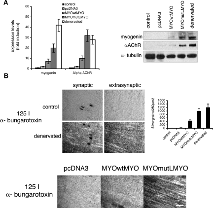
Figure 2.
MyogHCE controls AChRs extrasynaptic expression during postnatal muscle maturation. (A) Measurements of the myogenin and AChR α transcripts by qRT-PCR and immunoblot in leg muscle of 2-mo-old mice (control), after 48 h from denervation (denervated), or electroporated with constructs carrying myogenin cDNA driven by 1.1-kb wild-type myogenin promoter (MYOwtMYO) or mutant at myogHCE (MYOmutLMYO), or an empty pcDNA3 vector. Statistical significance of AChR α transcripts measurements in mutant MYOmutLMYO and in the denervated is P > 0.05. (B) The images show the AChRs density visualized by autoradiography of longitudinal sections from tibialis anterior muscle innervated (control) or 48-h denervated muscle in synaptic and extrasynaptic fiber segments (3 mm from the synaptic junctions) (top) or in extrasynaptic fiber segments (3 mm from the synaptic junctions) of tibialis anterior muscle electroporated with an empty pcDNA3 vector, myogenin cDNA driven by the wild-type myogenin promoter (MYOwtMYO) or the mutant at myogHCE (MYOmutLMYO) incubated with 125I-α-bungarotoxin (bottom). The sections were subsequently covered with a monolayer of photographic emulsion and after exposure and development silver grains were counted in 250-μm2 fields along the length of the sections, 3 mm on each side of the synapse (graph). The areas of high grain density in the left top panels correspond to the end-plate region of the fibers. Bar, 400 μm.

