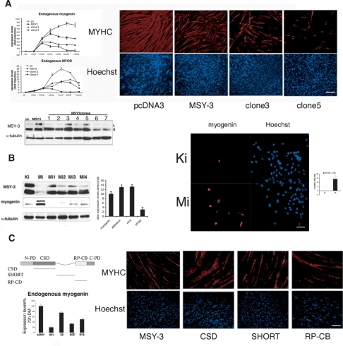
Figure 4.
Evidence MSY-3 inhibits myogenesis. (A) MSY-3 transfections of C2C12 myogenic cells. Expression during myogenic differentiation of endogenous myogenin and MyoD measured by qRT–PCR of mock-transfected (pc) and a MSY-3-transfected (MSY-3) C2C12 multiclonal populations, and two MSY-3-transfected clones (3 and 5). Immunoblot of extracts from MSY-3-overexpressing C2C12 clones blotted with ZONAB (MSY-3 Ab) and α-tubulin control. Two MSY-3 isoforms, short (S) and long (L). Myosin heavy chain Ab marks differentiation (left) and Hoechst (right) stains of nuclei of the same C2C12 multiclone populations and clones 3, 5 at 72 h in DM (full differentiation). Bar, 400 μm. (B) Effects of MSY-3 knockdown by siRNA. The immunoblot shows the two isoforms of MSY-3 protein in extracts from C2C12 in GM, transfected with a pool of four interfering oligos (MiSmart) against MSY-3; or single oligos (Mi1, Mi2, Mi3, Mi4), compared with control oligos (KiSmart). Levels of myogenin are reported and normalized relative to α-tubulin. Left graph shows the ratio of myogenin, MyoD, α AChR subunit, and MCK between the C2C12 in GM blocked with MSY-3 siRNA (MiSmart) or with control siRNAs (KiSmart). GAPDH was used to normalize. Anti myogenin Ab (left panel) and Hoechst (right) staining of control siRNA (KiSmart) or MSY-3 siRNA (MiSmart). Bar, 200 μm. Graph quantifies myogenin positive C2C12 nuclei in growth medium. (C) Mutational analysis of MSY-3 protein. C2C12-transformed cell lines overexpressing mutated forms of HA-tagged MSY-3 protein were derived. In Supplemental Figure 10A expression of MSY-3-HA tagged in MSY-3 wild-type and mutant polyclone populations is shown. Domain structure of MSY-3 protein and mutated forms derived N-PD (N-terminal proline-rich domain), CSD (cold-shock domain), SHORT (splicing alternative domain), RP-CB (arginine- and proline-rich conserved domain), C-PD (C-terminal praline-rich domain). The graph indicates the expression level of endogenous myogenin in the clones overexpressing the wild-type MSY-3 protein, and the mutated forms in the CSD, SHORT, and the RP-CB domain. Myosin heavy chain Ab marks differentiation (top) and Hoechst (bottom) stains nuclei of the same C2C12 multiclone populations at 72 h in DM (full differentiation). Bar, 400 μm.

