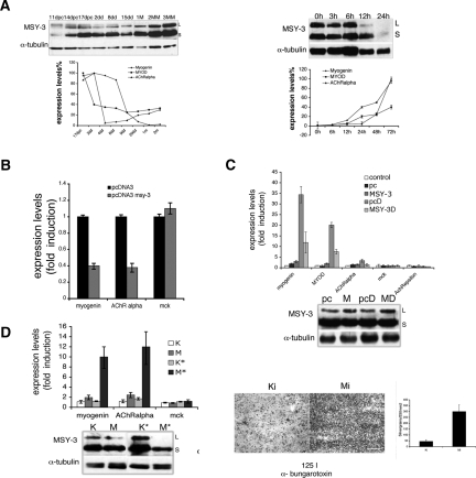
Figure 6.
MSY-3 in adult muscle innervation. (A) Expression of MSY-3 protein and myogenin, MyoD and AChR α subunit RNA in muscle at different times of maturation (immunoblot and graph, left) and after denervation by sciatic nerve transection (immunoblot and graph, right). (B) Expression of myogenin and AChR α subunit genes after electroporation in tibialis anterior and quadriceps muscle of young mice (14 d postnatal) of the control (pcDNA3) and MSY-3 (pcDNA3-MSY-3) constructs evaluated by qRT–PCR. (C) Expression of myogenin, MyoD, AChR α subunit, MCK, and AChR ε in adult (2 mo) tibialis anterior muscle (control) electroporated with pcDNA3 (pc); electroporated with MSY-3 (MSY-3); electroporated with the same constructs and later denervated by sciatic nerve transection (pcD and MSY-3D). Expression was evaluated by qRT–PCR and immunoblot (MSY-3). (D, left) Expression of myogenin, AChR α subunit, and MCK in tibialis anterior muscle of adult mice (2 mo) elecroporated with a pool of four interfering oligos (Mi), which blocks MSY-3 or with control oligos (Ki), coinjected with a nuclear GFP-expressing plasmid (Mi* and Ki*) in order to dissect only the injected area and evaluated by qRT–PCR. The immunoblot measures MSY-3 expression. (Right) AChRs density in extrasynaptic regions (3 mm from synaptic junctions) visualized by autoradiography of longitudinal sections from Ki* or Mi* electroporated tibialis anterior muscle of adult mice (2 mo old) and incubated with 125 I-α-bungarotoxin. Bar, 200 μm.

