Figure 3.
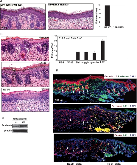
Exogenous noggin or SHH, but not by Wnt3a, can rescue hair development in the absence of epithelial laminin-511. (A) Hematoxylin-eosin staining of skin harvested 2 wk following hair reconstitution assay using either wild-type (WT) or lama5−/− (null) E16.5 keratinocytes combined with wild-type dermal cells. (Right) Quantification of hair follicles in both conditions. (B) Full thickness E16.5 lama5−/− dorsal skin was incubated overnight with purified Wnt3a protein, Shh adenovirus (SHH), or noggin and analyzed by hematoxylin-eosin staining 9 d after grafting to nude mice. (Top right) Quantification of hair follicles under six different conditions for hair restoration experiments, including PBS control. (C) Level of β-catenin in E16.5 lama5−/− skin after overnight treatment with indicated quantity of purified Wnt3a (in ng/mL) demonstrating the activity of recombinant Wnt3a. (D) Immunofluorescence microscopy of adjacent frozen sections confirming hair follicle formation in absence of laminin-511 in grafted skin treated with noggin. Laminin-511 negative hair follicles (arrows) were identified as clusters of cells (nuclear antibody, DAPI) showing positive keratin 17 and perlecan expression, and negative Pecam expression, distinguishing them from blood vessels. The expression of laminin-511 was visualized by laminin α5 pAb (L511); white dots indicate the border between graft and host skin.

