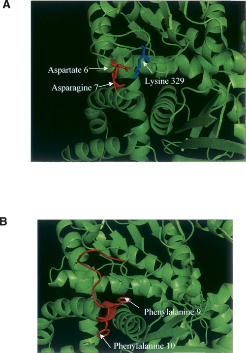
Figure 6.
(A) Close-up of an N-terminal interaction in a domain from 1HWY, a domain with bound glutamate, NADH, and GTP. Highlighted in red are aspartate 6, and asparagine 7, and the prominent lysine 329 (in blue) of the coenzyme-binding domain, which are all within 3.5 Å of each other. (B) PyMOL (DeLano Scientific) cartoon displaying a close-up of the N-terminal of bovine GDH (1HWY), and in particular, amino acids 1–10 in red with the F9 and F10 in stick form. These phenylalanine residues lie on either side of the third helix of the glutamate-binding domain, and the peptide bond between residues 10 and 11 is the site of cleavage by chymotrypsin.

