
Fig. 3.
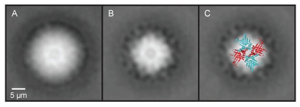
Transmission electron microscopy image reconstruction of native mitochondrial RNA-binding protein (MRP) 1/MRP2 complexes isolated from Trypnaosoma brucei. (A) View of the MRP1/MRP2 complex prior to the RNase treatment. Clearly visible in the structure is the central hole in the cavity of the MRP1/MRP2 heterotetramer complex. (B) View of the same complexes after the RNase treatment. Comparison of the RNase-treated and non-treated complexes indicates that the RNA does not bind in the central cavity but on the surface and/or the outside of the complex. (C) Superimposition of the crystal structure on an averaged electron microscopy picture of the MRP1/MRP2 complex.