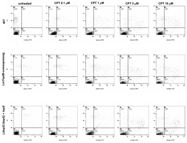
Figure 8.
PS externalization as a consequence of CPT exposure; WT, LmTopIB-overexpressing and rescued (ΔtopS/ΔtopS)+topS strains were treated with different concentrations of CPT for 24 h and analyzed for PCD. Dead cells were excluded by PI incorporation. Dot plots are representative of three independent assays.

