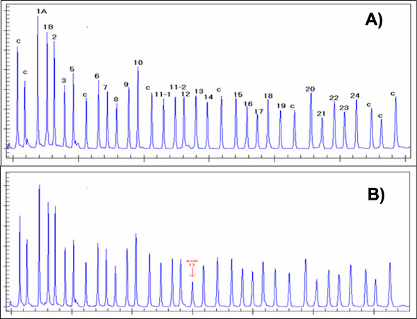
Figure 1.
Example of a deletion in an MLPA assay. Panels A and B show pherograms corresponding to the electrophoresis of an MLPA assay. In the Y-asix are depicted the intensity signals (peak heights) for each probe that are depicted in the X-axis according to their length (probe size). Peaks marked with a C correspond to control probes and peaks numbered from 1 through 24 correspond to region-specific probes. Panel A corresponds to a normal individual, while panel B corresponds to an individual with a deletion at probe #13 as visible by the reduced peak intensity in this pherogram.

