Figure 2.
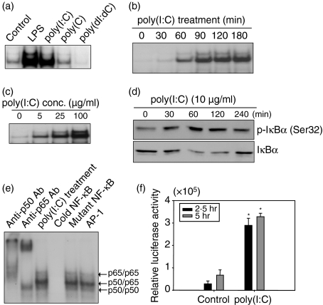
Nuclear factor-κB (NF-κB) activation with poly(I:C) stimulation in astrocytes. (a) Cells were treated with 100 ng/ml lipopolysaccharide (LPS), 25 μg/ml poly(I:C), 25 μg/ml poly(C) or 25 μg/ml poly(dI:dC) for 1 hr or left untreated. The NF-κB activation was analysed by electrophoretic mobility shift assay. (b) NF-κB activation increased in a time-dependent manner. Cells were stimulated with 25 μg/ml poly(I:C) for the indicated times. (c) NF-κB activation increased in a dose-dependent manner. Cells were treated with the indicated doses of poly(I:C) for 1 hr. (d) Cells were incubated with 10 μg/ml poly(I:C). After stimulation, cells were lysed by sonication and the lysates were analysed by Western blot using antibodies to p-IκBα or IκBα. (e) Antibody supershift assays were performed by preincubating poly(I:C)-treated nuclear extracts from astrocytes with the indicated antibodies. A 100-fold excess of unlabelled oligonucleotide (cold NF-κB), mutant oligonucleotide (mutant NF-κB) or non-specific oligonucleotide (AP-1) was used for competition assays. The data are representative of results obtained from three different experiments. (f) Luciferase reporter gene assay was performed. Fetal astrocytes were cotransfected with 0·05 μg/ml of pIL6-κB-Luc plasmid and pCMV-β-galactosidase vector. After 24 hr, cells were stimulated with poly(I:C). The luciferase activity was measured by a Luciferase Reporter Assay System. Luciferase activity is normalized for transfection efficiency with the β-galactosidase activity. *P< 0·05, significantly different from controls at the same treated time.

