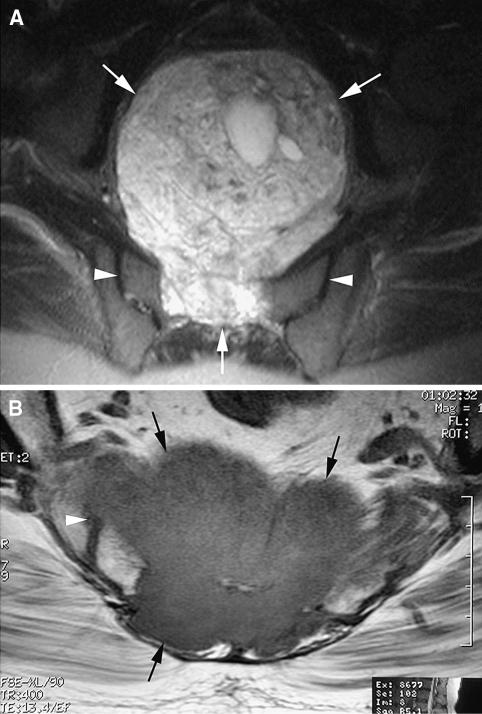
Fig. 1A–B.
(A) An axial fat-suppressed T2-weighted fast spin echo MR image shows a sacral chordoma with a large anterior soft tissue extension (arrows) but no involvement of the sacroiliac joints (arrowheads). (B) An axial T1-weighted spin echo MR image shows a sacral chordoma (arrows) with invasion of the right sacroiliac joint (arrowhead).

