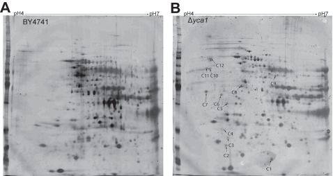
Figure 5. Representative 2D gels of BY4741 and Δyca1 proteomes.
(a) BY4741 and (b) Δyca1 cells separated along pH 4-7 IPG strips. The BY4741 gels were relatively over-stained to expose yca1 inducible protein species in the Δyca1 gel. Arrowheads depict spots that appear unique to Δyca1 lysates. Alphanumeric designation corresponds with identities summarized in table 1.

